
Orthopantomography or panoramic radiography is a radiological examination that allows you to study the teeth and the maxillary and mandibular alveolar bones. This type of radiography allows you to obtain a general and global vision of the whole mouth.
In Nart Dental Clinic we have the latest technology in the world of dental X-rays. Equipment to make: bitewing, apical radiography, CT or 3D radiography, orthopantomography and teleradiography.
When is it necessary?
Orthopantomography is used in Nart Dental Clinic to identify dental malformations such as included teeth, or bone pathology such as cysts and tumors. It also allows us to study the position of wisdom teeth and periodontal disease.
It is true that it is not as accurate in identifying carious lesions. When we are going to make a first visit to a patient it gives us an overview of the patient’s state of health. This test is sometimes complemented with other more specific radiographs in order to make a correct diagnosis.
How to perform an orthopantomography?
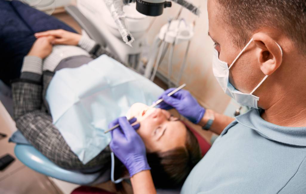
Orthopantomography is a very simple radiological test to perform. It does not require any previous preparation by the patient. The only thing that is recommended is to remove all metal objects such as earrings, necklaces, glasses, tongue piercing and removable dentures.
In Nart clinic in Barcelona is a widely used radiography that helps us to understand the dental situation of the patient and guides us to make a correct dental treatment.
It is a non-invasive examination, it does not cause any pain or discomfort.
Process of an orthopantomography

The execution time is about 20 seconds and once the examination is completed, no special care is required.
- Preparation of the patient: Before performing the test, a protective vest will be placed over the patient which serves to decrease the minimum radioactive levels, then the patient will be asked to remove all metallic objects.
- Positioning of the patient: Next, the patient will be positioned in front of the apparatus so that the orthopantomography will be successful. Technically, the patient is positioned standing in front of the radiographic apparatus and has to bite a plate in order to maintain the correct position and keep the head still.
- Use of the X-ray machine: Once the patient is in position and the equipment has been correctly adjusted, the X-ray exposure is performed. It is important that the patient remains still during this process to avoid distortions in the image.
- Image capture: The machine will create a panoramic image of the mouth and take care of processing it to improve its quality and clarity.
- Image interpretation: Finally, the dentist examines the orthopantomogram to identify any abnormalities, such as cavities, fractures, cysts, tumors or other dental or maxillofacial conditions.
Contraindications
However, being a radiological technique, it exposes the patient to very low doses of ionizing radiation. Like all radiological examinations, therefore, orthopantomography is contraindicated in pregnant patients.
The collaboration required between patient and operator makes the technique difficult to perform in children under 6 years of age and in all patients with psychiatric disorders or neurological pathologies.


